We take the ocular health of our patients seriously.

Optomap ultra-widefield retinal imaging is our standard of care and will be part of your comprehensive eye exam. This device helps you and your eye doctor make informed decisions about your eye health and overall well-being. Combining your eye doctor’s expertise and the Optomap technology, Optos brings your eye exam to life.


Ultra-Widefield Retinal Imaging Will Reveal:

  • Ocular Melanoma (Cancer)
  • Diabetic Retinopathy (DR)
  • Age-Related Macular Degeneration (AMD)
  • Glaucoma (Increased Eye Pressure)
  • Hypertension (High Blood Pressure)

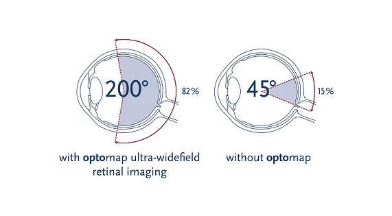
Optomap captures more than 80% of your retina.

Optomap captures more than 80% of your retina.

Examining the retina is challenging and requires looking through your pupil to examine the back of your eye. Traditional viewing methods can be effective, but are more difficult to perform and are carried out manually without any digital record. Optomap captures more than 80% of your retina in one image where traditional methods typically reveal only 10-15% of your retina at one time. This technology enhances our ability to detect the earliest signs of disease that appear on your retina.  An optomap only takes seconds to perform, and is not painful, and typically does not require dilation. However, a doctor may decide dilation is still needed.

What is your Retina?

What is your Retina?

The retina is a delicate lining at the back of the eye similar to film in a camera. Light strikes the retina through the lens in your eye and produces a picture which is then sent to the brain, enabling you to see. Your retina is the only place in the body where blood vessels can be seen directly. This means, in addition to eye conditions, signs of other diseases (such as stroke, heart disease, hypertension and diabetes) can also be seen in the retina. Early detection is essential so proper treatment can be administered.


An unhealthy retina cannot send clear signals to the brain which can result in impaired vision and blindness. Most retinal conditions and other diseases can be successfully treated with early detection. Without a comprehensive eye exam, you may not be aware of a potential problem. You may even see clearly now, but because the retina has no nerve endings, you would not feel pain – a symptom which prompts you to see a doctor.

Retinal Detachment

When the retina detaches, it is lifted or pulled from its normal position. If not promptly treated, a retinal detachment can cause permanent vision loss. Anyone can get a retinal detachment; however, they are far more common in nearsighted people, those over 50, those who have had significant eye injuries, and those with a family history of retinal detachments.


What are the different types of retinal detachments?

Rhegmatogenous [reg-ma-TAH-jenous] – A tear or break in the retina causes it to separate from the retinal pigment epithelium (RPE), the pigmented cell layer that nourishes the retina, and fill with fluid. These types of retinal detachments are the most common.


Tractional – In this type of detachment, scar tissue on the retina’s surface contracts and causes it to separate from the RPE. This type of detachment is less common.


Exudative – Frequently caused by retinal diseases, including inflammatory disorders and injury/trauma to the eye. In this type, fluid leaks into the area underneath the retina (subretina).


Early detection of all of these diseases and conditions mean successful treatments can be administered which reduces the risk to your sight and health.
FAQs

  • An unhealthy retina cannot send clear signals to the brain which can result in impaired vision and blindness. Most retinal conditions and other diseases can be successfully treated with early detection. Without a comprehensive eye exam, you may not be aware of a potential problem. You may see clearly, and because the retina has no nerve endings, you may not feel any pain, a symptom which may otherwise prompt you to see a doctor.

  • Your retina is the only place in the body where blood vessels can be seen directly. This means, in addition to eye conditions, signs of other diseases (such as stroke, heart disease, hypertension and diabetes) can also be seen in the retina. Early detection is essential so treatments can be administered.


    Diabetic Retinopathy (DR).

    Diabetes affects the eyes and the kidneys and is a leading cause of blindness. Retinopathy occurs when diabetes damages the tiny blood vessels inside the retina.


    Age-related Macular Degeneration (AMD).

    The center of the retina (the macula) can become diseased when we get older. The results in alterations to our fine central vision making daily activities difficult.


    Glaucoma (Increased Eye Pressure).

    Glaucoma causes damage to the optic nerve and almost always develops without symptoms.


    Hypertension (High Blood Pressure).

    Increased pressure can result in changes to blood vessels in the eye, increasing the risk of cardiovascular disease (stroke or heart disease).


    Ocular Melanoma

    Ocular melanoma is often lethal, but thankfully, a very rare disease. There are other types of eye cancers, but melanoma is the most common. Choroidal melanomas are the most common site comprising 85% of cases. Other sites include ciliary body, iris, and conjunctiva. Current research is also showing that ocular melanoma is very different from normal skin melanoma.

  • Examining the retina is challenging. Your doctor looks through your pupil to examine the back of your eye. Traditional viewing methods can be effective, but difficult to perform and are carried out manually without any digital record.

  • The optomap ultra-wide digital retinal imaging device captures more than 80% of your retina in one image. Traditional methods typically reveal only 10-15% of your retina at one time. The unique optomap ultra-widefield view enhances your eye doctor’s ability to detect even the earliest signs of disease that appears on your retina. Seeing most of the retina at once allows your eye doctor more time to review your images and educate you on your eye health. Numerous clinical studies have demonstrated the power of optomap as a diagnostic tool.

  • Your doctor will advise you based on your individual circumstances, but the general recommendation is that you have an optomap every time you have an eye exam. This will ensure you have a digital record of your retinal health on file which can be compared for changes over time.

  • Many vision problems begin at an early age, so it’s important for children to receive proper eye care from the time they are infants.

  • An optomap only takes seconds to perform, and is not painful, and typically does not require dilation. However, your eye doctor may decide dilation is still needed.

  • Optos was founded over 20 years ago by Douglas Anderson after his then five-year-old son, Leif, went blind in one eye when a retinal detachment was detected too late. Routine exams were uncomfortable, especially for a child, which made it impossible for the doctor to conduct a complete exam and view the entire retina. Douglas set out to develop a patient-friendly retinal imaging product that encompassed a digital ultra-widefield image of the retina, easily.


    “With the optomap exam, Leif’s detached retina might have been detected in time to be properly treated. His eye could have been saved.” – Douglas Anderson

(function(){var s = document.createElement("script");s.setAttribute("data-account","nQM1O9ax3C");s.setAttribute("src","https://cdn.userway.org/widget.js");document.body.appendChild(s);})();Enable JavaScript to ensure website accessibility